Test Your Eyes
READING HRCT SCANS
Refine your skills by viewing HRCT scans of the lungs of patients with suspected IPF and identify specific features found in images that are consistent with a radiological diagnosis of usual interstitial pneumonia (UIP), probable UIP, indeterminate for UIP, or those that are suggestive of an alternative diagnosis.
HRCT SCAN (1 OF 4)
Images courtesy of and used with permission from Jonathan Goldin, MD, PhD.
WHAT RADIOGRAPHIC FEATURES ARE PRESENT IN THE HRCT SCANS SHOWN HERE?
Choose all that apply.
Honeycomb formation
Subpleural, basal predominance
Micronodules
Air trapping
Reticular abnormality
Images courtesy of and used with permission from Jonathan Goldin, MD, PhD.

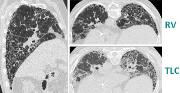
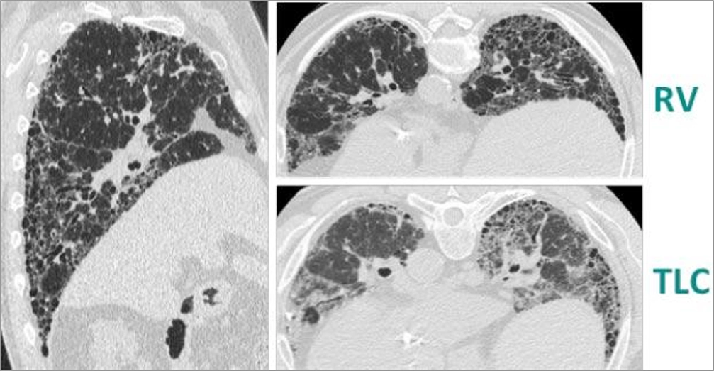
The answer is: honeycomb formation, subpleural, basal predominance and reticular abnormality. The radiographic features seen in these HRCT scans meet the criteria for UIP. Usual interstitial pneumonia is spatially and temporally heterogeneous with variant areas of normal lung, interstitial inflammation, fibrosis, and honeycombing.
In the left sagittal image, there is clear evidence of peripheral, lower lobe predominance of reticulation with honeycomb formation. In the TLC and RV scans on the right, there is honeycomb formation. Finally, the RV scan shows a uniform decrease in attenuation across functioning parenchyma, with no evidence of air trapping.
Images courtesy of and used with permission from Jonathan Goldin, MD, PhD.

The answer is: honeycomb formation, subpleural, basal predominance and reticular abnormality. The radiographic features seen in these HRCT scans meet the criteria for UIP. Usual interstitial pneumonia is spatially and temporally heterogeneous with variant areas of normal lung, interstitial inflammation, fibrosis, and honeycombing.
In the left sagittal image, there is clear evidence of peripheral, lower lobe predominance of reticulation with honeycomb formation. In the TLC and RV scans on the right, there is honeycomb formation. Finally, the RV scan shows a uniform decrease in attenuation across functioning parenchyma, with no evidence of air trapping.
HRCT SCAN (2 OF 4)
Images courtesy of and used with permission from Jonathan Goldin, MD, PhD.
THE IMAGES SHOWN IN THESE HRCT SCANS ARE:
Choose the correct answer:
Indicative of a UIP pattern
Indicative of a probable UIP pattern
Pattern indeterminate for UIP
Indicative of an alternative diagnosis
Images courtesy of and used with permission from Jonathan Goldin, MD, PhD.
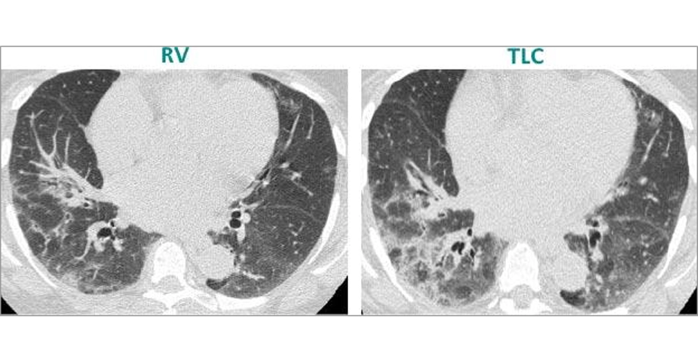
The answer is: alternative diagnosis. The lower lobe predominance, peribronchovascular disease, extensive ground glass abnormality with peripheral reticulation and air trapping clearly shown in these HRCT scans are all features that suggest a pattern other than UIP.
Images courtesy of and used with permission from Jonathan Goldin, MD, PhD.
The answer is: alternative diagnosis. The lower lobe predominance, peribronchovascular disease, extensive ground glass abnormality with peripheral reticulation and air trapping clearly shown in these HRCT scans are all features that suggest a pattern other than UIP.
HRCT SCAN (3 OF 4)
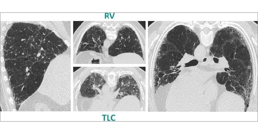
Images courtesy of and used with permission from Jonathan Goldin, MD, PhD.
THE PRESENCE OF WHICH OF THE FOLLOWING FEATURES WOULD SUGGEST A DIAGNOSIS OF PROBABLE UIP IN THIS PATIENT?
Choose all that apply:
Lower lobe, subpleural predominance
Honeycomb formation
Air trapping
Reticular abnormalities
Profuse micronodules
Images courtesy of and used with permission from Jonathan Goldin, MD, PhD.
The answer is: lower lobe, subpleural predominance and reticular abnormalities. While there is clear lower lobe, subpleural predominance with reticular abnormalities in this patient’s HRCT scans, there is no evidence for true honeycomb cysts which would be suggestive of UIP. The TLC and RV scans show decreased attenuation and no significant evidence of air trapping. In addition, there is no evidence of micronodules which would suggest an alternative diagnosis to UIP. Taken together, this pattern is consistent with probable UIP.
Images courtesy of and used with permission from Jonathan Goldin, MD, PhD.
The answer is: lower lobe, subpleural predominance and reticular abnormalities. While there is clear lower lobe, subpleural predominance with reticular abnormalities in this patient’s HRCT scans, there is no evidence for true honeycomb cysts which would be suggestive of UIP. The TLC and RV scans show decreased attenuation and no significant evidence of air trapping. In addition, there is no evidence of micronodules which would suggest an alternative diagnosis to UIP. Taken together, this pattern is consistent with probable UIP.
HRCT SCAN (4 OF 4)
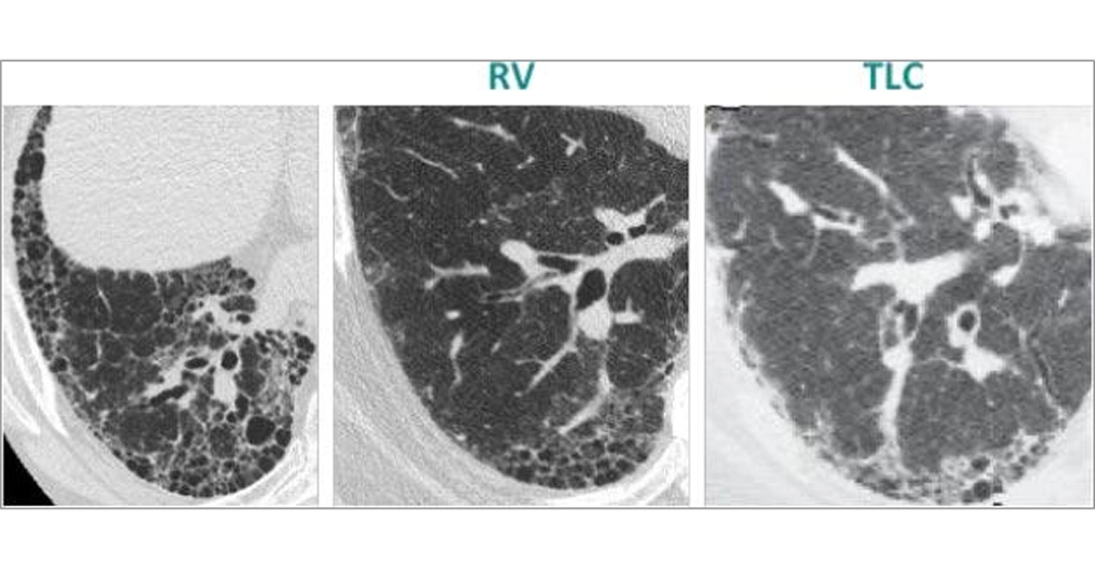
Images courtesy of and used with permission from Jonathan Goldin, MD, PhD.
THE IMAGES SHOWN IN THESE HRCT SCANS ARE:
Choose the correct answer:
Indicative of a UIP pattern
Indicative of a probable UIP pattern
Pattern indeterminate for UIP
Indicative of an alternative diagnosis
Images courtesy of and used with permission from Jonathan Goldin, MD, PhD.
The answer is: indicative of a UIP pattern. These images show clear evidence of honeycomb formation, which is central to the diagnosis of UIP. In addition to honeycomb change, the TLC and RV scans show lower lobe predominance, based on the presence of the inferior pulmonary vein. In the RV scan, there is uniform decreased attenuation and no evidence of air trapping. Therefore, the radiological diagnosis for this patient is UIP.
Images courtesy of and used with permission from Jonathan Goldin, MD, PhD.
The answer is: indicative of a UIP pattern. These images show clear evidence of honeycomb formation, which is central to the diagnosis of UIP. In addition to honeycomb change, the TLC and RV scans show lower lobe predominance, based on the presence of the inferior pulmonary vein. In the RV scan, there is uniform decreased attenuation and no evidence of air trapping. Therefore, the radiological diagnosis for this patient is UIP.
Your score
50%
You answered correctly 2 out of 4 questions.
Images courtesy of and used with permission from Jonathan Goldin, MD, PhD.