Case 3: 40 Year Old Female

FEMALE, 40 YEARS OF AGE
Patient referred to the lung disease clinic after reporting shortness of breath.
Physical Exam & Review
of Symptoms

Case Review
- Female, 40 years of age.
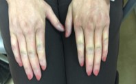
- Presents with shortness of breath and Raynaud's Phenomenon.

Key Findings
- Has restrictive pattern on PFTs
- HRCT-NSIP
- Skin thickening
- Serology positive for ANA and Scl - 70
Patient history
Past Medical/Surgical History
- Melanoma
- Bilateral breast reduction
- Poor skin healing
- Mouth tightening

- Mother: Arthritis, hypertension (HTN), high cholesterol, Diabetes
- Father: HTN, MI, Arthritis
- Never Smoker
- Rare ETOH
- Hispanic

Medications Prescribed Prior to Initial Visit
- Non-steroidal anti-inflammatory drug (NSAID)
- Non-NSAID analgesic
- Proton pump inhibitor
HRCT
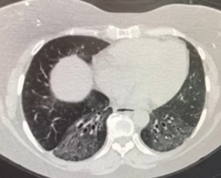
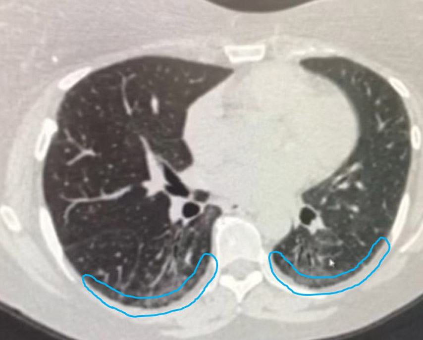
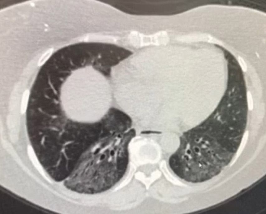
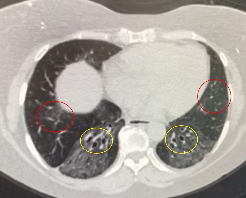
Your radiologist tells you the CT scan has the following features:
- [RED] Bilateral ground glass opacities
- [BLUE] Subpleural sparing
- [YELLOW] Traction bronchiectasis
Case study courtesy of and used with permission from Marilyn Glassberg, MD.
Physical Exam & Review
of Symptoms

Physical Exam
- BP: 150/92 mmHg, HR: 73, RR: 18
- Temperature: 98.6
- Weight: 189 lbs
- Height: 5’5”
- SpO2: 94%
- Raynaud’s Phenomenon

PFTs
- FVC 3.75 (63%)
- FEV1 3.08 (67%)
- FEV1/FVC Ratio 87%
- TLC 5.26 (53%)
- VC 3.70 (63%)
- RV 1.65 (25%)
- DLCO 24.4 (43%)
Serology Testing

Serologies
- lgG subclasses - normal
- dsDNA panel - negative
- Cyclic citrullinated peptide (CCP) - negative
- Rheumatoid factor (RF) - negative
- Sjögren's syndrome-A (anti-Ro) (SS-A) - negative
- Sjögren's syndrome-B (anti-La) (SS-B) - negative
- Anti-nuclear antibody (ANA) - 1:640 speckled
- Scleroderma-70 (SCL-70; Topoisomerase I) - positive
- TPO (thyroid peroxidase) 20.7 (high)
- Centromere B - negative
- Myositis panel — negative
- JO-1 - negative
- Aldolase — negative
- CK (creatine kinase) - normal
ANA, Anti-nuclear antibody; CCP, Cyclic citrullinated peptide; HP, hypersensitivity pneumonitis; RF, rheumatoid factor; SS-A, Sjögren's syndrome-A (anti-Ro); SS-B, Sjögren's syndrome-B (anti-La); SCL-70, Scleroderma-70 (topoisomerase I).
Case study courtesy of and used with permission from Marilyn Glassberg, MD.
References
- Mayo Clinic Laboratories website. Available at https://www.mayocliniclabs.com/test-catalog/Clinical+and+Interpretive/81359. Accessed April 22, 2019.
- Visscher D and Myers J. Histologic spectrum of idiopathic interstitial pneumonias. Proc Am Thorac Soc. 2006;3(4):322-9. doi: 10.1513/pats.200602-019TK.
- du Bois RM. Fibroblastic foci: time to be counted? Chest. 2006;130(1):3-5. doi: 10.1378/chest.130.1.3.
- Jones MG, Fabre A, Schneider P, et al. Three-dimensional characterization of fibroblast foci in idiopathic pulmonary fibrosis. JCI Insight. 2016;1(5):e86375. doi: 10.1172/jci.insight.86375.
Which is the correct
Diagnosis?

Antisynthetase Syndrome

Rheumatoid Arthritis

Interstitial Lung Disease Associated with Systemic Sclerosis

IPF
Correct!

Interstitial Lung Disease Associated with Systemic Sclerosis
Reynaud‘s phenomenon, NSIP on HRCT, positive ANA and Scl-70 are consistent with systemic sclerosis.
Incorrect!

Antisynthetase Syndrome
While restrictive defects of PFTs, NSIP on HRCT, and Reynaud‘s phenomenon are consistent with antisynthetase syndrome, the serology test is not. Elevated levels of anti-J01 are consistent with antisythetase syndrome, but the serology in this patient is negative for anti-Jo 1.
Incorrect!
Idiopathic Pulmonary Fibrosis
HRCT is consistent with an NSIP pattern that would not be found in HRCT from patients with IPF.
Incorrect!
Rheumatoid Arthritis
While restrictive defects of PFTs and NSI P on HRCT are consistent with rheumatoid arthritis, the serology test and skin manifestation is not. Elevated levels of anti—RF or anti-CCP are consistent with rheumatoid arthritis, but the serology in this patient is negative for anti-RF.
Find out the answer

Interstitial Lung Disease Associated with Systemic Sclerosis
Reynaud‘s phenomenon, NSIP on HRCT, positive ANA and Scl-70 are consistent with systemic sclerosis.