Case 2: 67 Year Old Male

MALE, 67 YEARS OF AGE
Presents with 2-3 years history of cough and progressive dyspnea.
Physical Exam & Review
of Symptoms

Case Review
- Male, 67 years of age.
- Presents with 2-3 years history of cough and progressive dyspnea.
- No clinically relevant symptoms.

Key Findings
- Has restrictive pattern on PFTs
- Serologies - unremarkable
- HRCT - traction bronchiectasis; honeycombing
- Histopathology: microcystic honeycombing, patchwork distribution
Patient history
Past Medical/Surgical History
- GERD
- Seasonal allergies

- No Family History noted
- Ex-smoker, 20 pack-years
- No Drug use

Medications Prescribed Prior to Initial Visit
- Proton Pump Inhibitors
HRCT
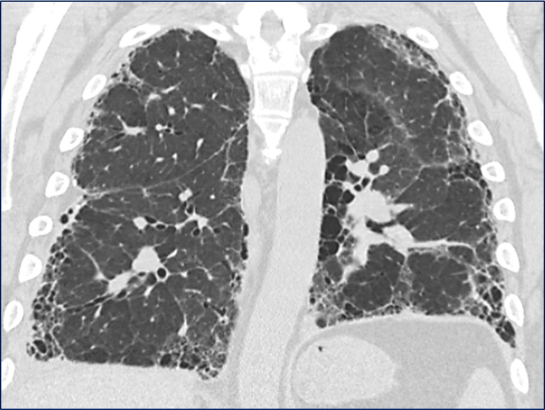
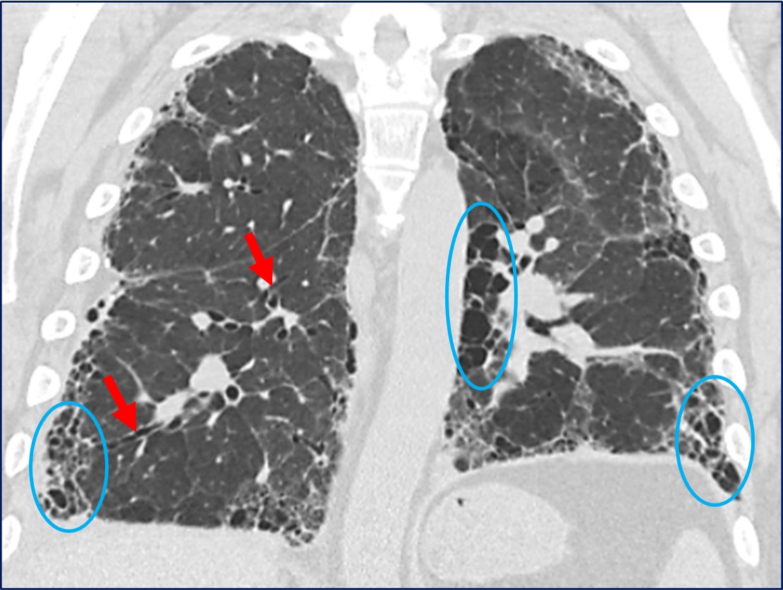
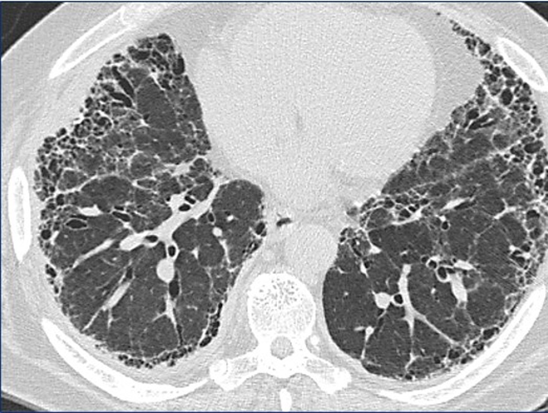
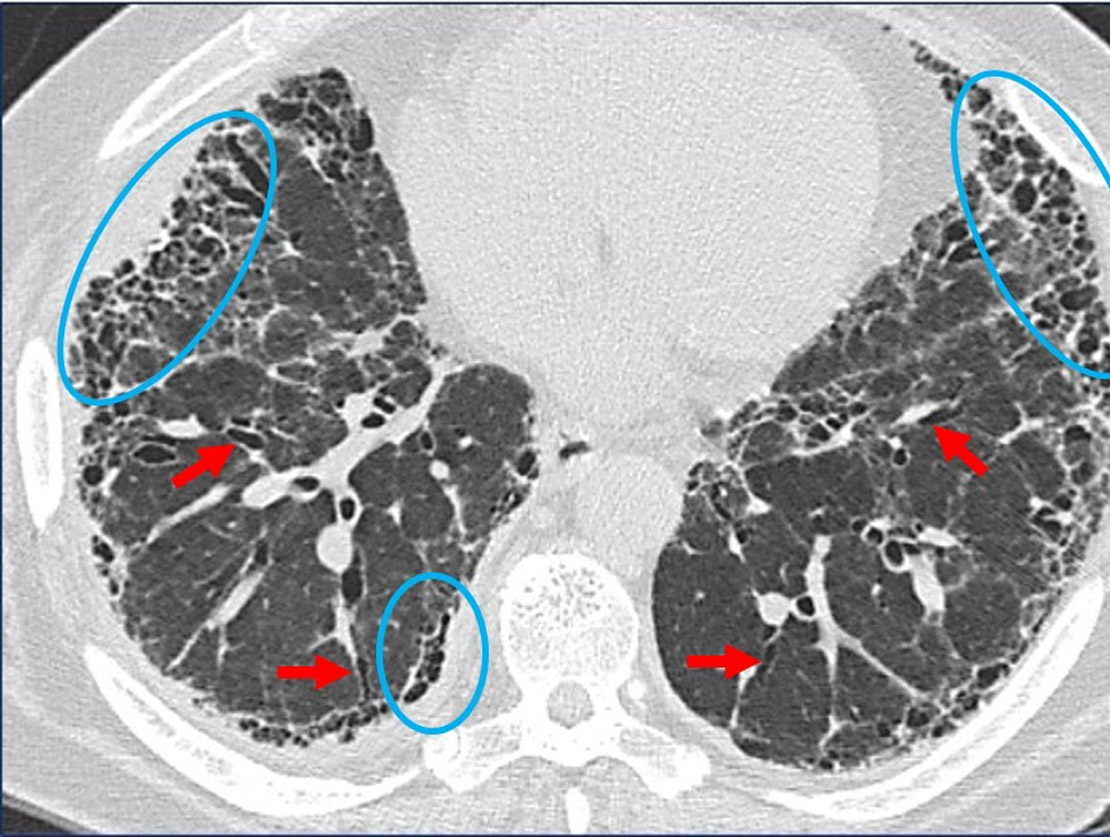
Your radiologist tells you the CT scan has the following features:
- ➔ Traction bronchiectasis
- Honeycombing
Case study courtesy of and used with permission from Robert Suh, MD.
Physical Exam & Review
of Symptoms

Physical Exam
- BP: 148/82 mmHg, HR: 75, RR: 18
- Temperature: 98.1
- Weight: 194 Ibs
- Height: 5’10”
- SpO2: 92%
- Well appearing
- Lungs: mid-late inspiratory crackles
- Digital clubbing

PFTs
- FVC 2.97 (67%)
- FEV1 2.64 (82%)
- FEV1/FVC Ratio 89%
- TLC 4.48 (67%)
- VC 3.48 (77%)
- RV 1.27 (47%)
- DLCO (73%)
Surgical lung biopsy

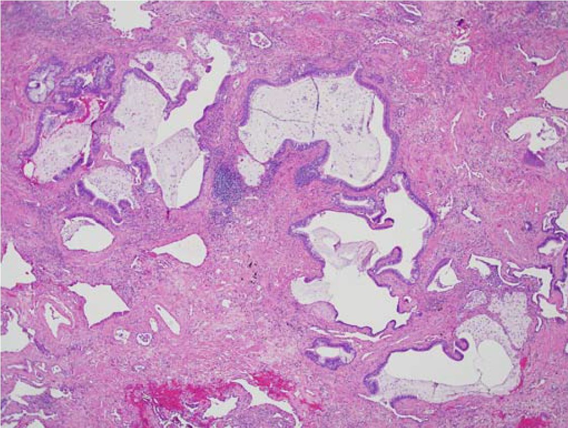
Image used with permission from Visscher D and Myers J. Proc Am Thorac Soc. 2006;3(4):322-9.
Low magnification image
Image used with permission from Visscher D and Myers J. Proc Am Thorac Soc. 2006;3(4):322-9.
Histopathology:
- High magnification: Fibroblast focus (arrows)
What are fibroblastic foci?
Fibroblastic foci -aggregates within the lung consisting of proliferating fibroblasts and myofibroblasts with a localized edge of spindle cells these indicate areas of active fibrosis.1-3
References
- Visscher D and Myers J. Proc Am Thorac Soc. 2006;3(4):322-9.
- Du Bois. CHEST. 2006;130(1):3-5.
- Jones et al. JCI Insight. 2016;1(5):e86375.
Pathology:
Low magnification
- Honeycomb change
- Columnar respiratory epithelium outline the cystic spaces
- Cystic spaces contain mucus and inflammatory cells
See additional note from pathologist:
NOTE: Additional sample shows patchy distribution of abnormalities
Serology Testing

Serologies
- Specific immunoglobulin panel - negative
- Cyclic citrullinated peptide (CCP) - negative
- Rheumatoid factor (RF) - negative
- Sjögren’s syndrome-A (Anti Ro) (SS-A) - negative
- Sjögren’s syndrome-B (anti-La) (SS-B) - negative
- Anti-nuclear antibody (ANA) - negative
- Scleroderma-70 (SCL-70; Topoisomerase 1) - negative
Case study courtesy of and used with permission from Gregory Cosgrove, MD.
Which is the correct
Diagnosis?

Sarcoidosis

Rheumatoid Arthritis

LAM

IPF
Correct!

Idiopathic Pulmonary Fibrosis
This is correct! The clinical and HRCT results are characteristics of IPF.
Incorrect!

Rheumatoid Arthritis
The patient had negative CCP and RF antibodies, both of which are associated with rheumatoid arthritis. Furthermore, the patient had no joint involvement or other signs and symptoms of rheumatoid arthritis.
Incorrect!
Lymphangioleiomyomatosis
A diagnosis of LAM is incorrect because it occurs exclusively in women. Furthermore, reduced RV and TLC suggests an alternative diagnosis. The cysts found in this patient‘s HRCT are consistent with lower-lobe honeycombing, not the small diffusely distributed cysts seen in LAM.
Incorrect!
Sarcoidosis
A diagnosis of sarcoidosis is unlikely because the HRCT shows lower lobe honeycombing. In sarcoidosis, nodules in the upper or middle lobes are common. Also, pathology does not show non-caseating granulomas, which is a key feature of sarcoidosis.
Find out the answer

Idiopathic Pulmonary Fibrosis
The clinical and HRCT results are characteristics of IPF.