Sarcoidosis
DIAGNOSIS
How is sarcoidosis diagnosed?
Diagnostic Tests
Workup should achieve 4 things:1
- Histologic confirmation of sarcoidosis
- Assessment of extent and severity of organ involvement
- Assessment of disease stability or likelihood to progress
- Determine if therapy will be beneficial to the patient
DIAGNOSTIC TOOLS:1
- Patient evaluation
- Histology and immunological screening
- Chest X-ray
- Used to determine stage of disease1
- Computed Tomography (CT) scans
PATIENT EVALUATION
A comprehensive evaluation for all patients should include:1
- Symptom evaluation
- Patient history (occupational and environmental exposure)
- Physical exam and routine eye exam
- Posteroanterior chest X-ray
- PFTs: spirometry and DLco
- Most likely to indicate functional impairment
- Can uncover both restrictive and obstructive defects
- Baseline assessment
- Instrumental to monitor improvement or deterioration of lung disease
SYMPTOMS
Pulmonary sarcoidosis is typically found by abnormal chest X-ray, often incidentally.9
- Typically bilateral mediastinal adenopathy if asymptomatic — if symptomatic, most commonly dyspnea, cough, nonspecific chest discomfort, crackles4
- If wheezing is present, this is often a sign of fibrosis3
Symptomatic patients display:
- Dyspnea, cough, chest pain1 :— 1/3 to 1/2 patients1
- Lung crackIes1 :— less than 1/5 patients1
- Clubbing is rare1
Other Symptoms4
- Fatigue
- Malaise
- Weakness
- Weight Loss
- Low-grade Fever
- Anorexia
- Symptoms depend on the site and degree of involvement, varying over time
- Can range from spontaneous remission to chronic indolent illness
- It is common for patients with sarcoidosis to not have presenting symptoms
LABORATORY AND PHYSICAL EXAMINATIONS
- Peripheral blood counts1
- White blood cells
- Red blood cells
- Platelets
- Serum chemistries1
- Calcium
- Liver enzymes
- Creatinine
- Blood Urea Nitrogen (BUN)
- Urine analysis1
- Electrocardiogram (ECG1)
- Tuberculin skin test1
- CT scans may be recommended in some patients pending results
HISTOLOGY
Common Histologic and Immunologic Findings:
Histologic: Transbronchial Lung Biopsy (TLB)
- Performed in presence of a compatible clinical picture1
- Should be cultured for fungi and mycobacteria to exclude other diagnoses4
- Mediastinoscopy may be favorable to TLB10
Immunologic Features:1
- Depression of cutaneous delayed-type hypersensitivity
- Heightened T helper cell type 1 (TH1) immune response at sites of disease
- Circulating immune complexes, along with signs of B cell hyperactivity, may also be found
CHEST X-RAY
Chest X-rays are used to determine the stage of disease:
Stage 0 - Normal chest X-ray1
Stage I - Bilateral hilar lymphadenopathy; may be accompanied by paratracheal adenopathy; parenchymal granulomas upon lung biopsy1
Stage II - Bilateral hilar adenopathy accompanied by pulmonary infiltration1
Stage III - Pulmonary infiltration without bilateral hilar lymphadenopathy1
Stage IV - Pulmonary fibrosis evidenced by honeycombing, hilar retraction, bullae, cysts, and emphysema1
CT SCANS
Typically only used in the following instances:1
- Atypical clinical and/or chest radiograph findings
- Detection of complications due to lung disease
- Normal chest radiograph, but clinical suspicion of disease
Classic Findings1
- Widespread small nodules with bronchovascular and subpleural distribution
- Thickened interlobular septae
- Architectural distortion
- Conglomerate masses
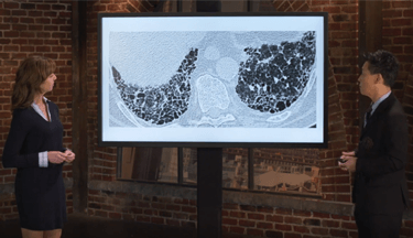
CT Scan: Classical Findings
The CT scans of 80 patients were evaluated by Abehsera and colleagues.11
47% - Bronchial Distortion11
29% - Honeycombing11
24% - Linear Fibrosis11
- Fibrotic sarcoidosis and bronchial distortion (marked by traction bronchiectasis and airway angulation), linear scarring, and honeycombing were found11
- Extent and type of fibrosis in sarcoidosis varies, which may account for the variety of clinical findings and outcomes3,11
- Although fibrosis is often upper- and middle-lobe predominant, the distribution of fibrosis may be somewhat pattern specific3
- In the study by Abehsera et al, honeycombing was observed more frequently in the upper lobes, and lower lung involvement was more common for diffuse linear fibrosis11
- Regular follow-up of patients with ground-glass opacities may be warranted, even when there are minimal pulmonary symptoms initially3
Less Common Findings by CT Scan
- Honeycombing1
- Cyst formation and bronchiectasis1
- Alveolar consolidation1