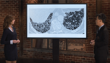
Occupational ILD - Diagnosis
DIAGNOSIS
CLINICAL PRESENTATION AND DIAGNOSTIC CONSIDERATIONS3
- The clinical, radiologic, and pathologic presentations of occupational ILDs are similar to non-occupational lLDs due to the lung’s general response to injury
- A thorough, lifetime, work, and environmental history is critical to identify potential occupational exposures
- Characteristic radiographic changes in combination with a work history is sufficient to make diagnosis
- Occupational ILD should be considered for any new ILD cases in the absence of a known cause
- Material safety data sheets should be obtained where possible
- Pulmonary function tests are useful to determine severity and pharmacologic treatment
PRESENTING SYMPTOMS
- Most common symptoms in patients with significant disease8
- Shortness of breath
- Cough
- Chest tightness
- Wheezing
- Patients with less significant disease will have no respiratory symptoms9
- Diagnosis is often made based on occupational history and routine chest X-ray (CXR)8
DIAGNOSIS REQUIRES:3
- History of exposure to a known ILD-inducing agent
- An appropriate latency period following exposure
- Consistent clinical disease course
- Pattern of physiologic and radiologic evidence of disease
- Exclusion of other known ILD-causing factors
- Lung biopsy is not needed if these tests come back positive, but recommended for atypical cases or cases arising from new or poorly understood causative agents
DIAGNOSTIC TESTS
- After an occupational history is collected, ILD is typically detected through use of radiologic imaging, usually a CXR10
- Spirometric testing, high-resolution computed tomography (HRCT), sputum analysis, and bronchoalveolar lavage are also recommended10
- The International Labor Office (ILO) provides guidelines for the systematic scientific classification of pneumoconiosis5
- The CXR is classified and scored on the following features:5
- Film quality5
- Rounded small opacities (diameter)5
- Irregular small opacities (width)5
- Profusion (concentration of small opacities in affected area of the lung)5
- Large opacities (longest dimension >10 mm)5
| Exposure11 | Imaging11 |
|---|---|
| Coal mine dust |
|
| Silica |
|
| Asbestos |
|
| Beryllium |
|